 08045816368


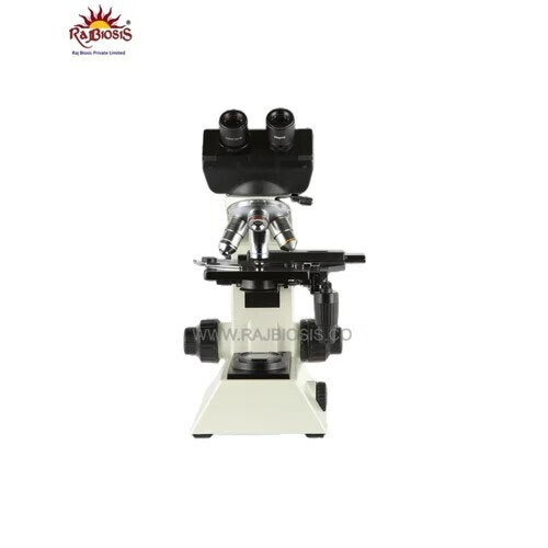
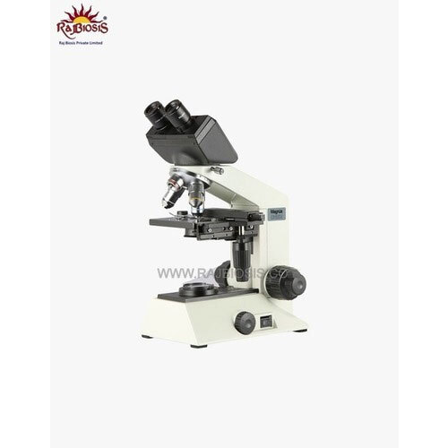
Research Trinocular Microscope
68000 INR/Piece
Product Details:
- Focus System Coarse and fine coaxial focusing system.
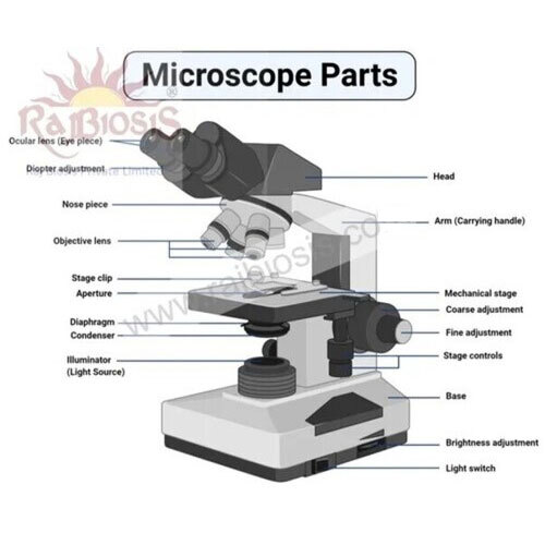
- View Head Trinocular head, inclined at 45°, rotatable 360°.
- Features Anti-fungal coating, metal stand, ergonomic design.
- Spare Parts Optional eyepieces, objectives, bulbs, fuse.
- Theory Brightfield and darkfield microscopy for biological and research applications.
- Drawtube Trinocular drawtube for simultaneous viewing and photomicrography.
- Sensor High-sensitivity CMOS digital camera (when attached, optional).
- Click to view more
X
Research Trinocular Microscope Price And Quantity
- 1 Piece
- 68000 INR/Piece
Research Trinocular Microscope Product Specifications
- Abbe condenser NA 1.25 with iris diaphragm.
- Approx. 380mm x 270mm x 180mm.
- Optional eyepieces, objectives, bulbs, fuse.
- LED/Halogen built-in, brightness adjustable.
- Brightfield and darkfield microscopy for biological and research applications.
- Built-in LED or Halogen 6V/20W adjustable intensity.
- Up to 30 fps (with attached camera accessory).
- Up to 1920 x 1080 pixels (for digital camera attachments).
- USB 2.0/3.0 interface for camera connectivity.
- 1920 x 1080 pixels (camera dependent).
- Anti-fungal coating, metal stand, ergonomic design.
- High-sensitivity CMOS digital camera (when attached, optional).
- Trinocular head, inclined at 45°, rotatable 360°.
- JPG, BMP, TIFF (with integrated camera system).
- 55 - 75mm adjustable.
- Coarse 30mm, fine 0.2mm per division.
- Mechanical stage 140 x 140mm; X-Y movement 75 x 50mm.
- Coarse and fine coaxial focusing system.
- Achromatic objectives: 4x, 10x, 40x (spring), 100x oil (spring).
- 40x to 1000x (with interchangeable objective lenses and eyepieces).
- 0.2mm/division.
- 30mm full range.
- Trinocular tube, 45° inclined, 360° rotatable.
- Full HD 1080p (camera dependent).
- Trinocular drawtube for simultaneous viewing and photomicrography.
- Standard focal distance 160mm (objective-tube length).
- Wide field WF10x/18mm (paired), optional WF15x/16x.
- Sturdy heavy metal base for vibration-free operation.
- Scratch-resistant hard-coated surface.
- Quadruple revolving nosepiece with positive click stop.
- Standard off-white/grey finish.
- Yes, on left eyepiece tube.
- AC 220V/110V, 50/60Hz.
- Supplied with dust cover.
- Medical research, biology, pathology and clinical laboratories.
Research Trinocular Microscope Trade Information
- 30 Piece Per Day
- 1-2 Days
Product Description
Experience the marvels of modern microscopy with the Research Trinocular Microscope-engineered for medical research, pathology, and biological labs. Featuring a lavish quadruple revolving nosepiece with positive click stops, scratch-resistant hard-coated mechanical stage, and a sturdy, heavy metal base for vibration-free operation. The latest camera compatibility allows access to high-resolution imaging up to 1920x1080 pixels. With ergonomic design, anti-fungal coating, and valuable dual illumination (LED/Halogen), this microscope meets the premium needs of research professionals. Shop now for essential innovation in laboratory observation!
Key Features & Applications of Research Trinocular Microscope
Unlock superior performance with the Research Trinocular Microscope's quadruple nosepiece, trinocular head, and advanced illumination options. Designed for seamless use in medical research, biology, pathology, and clinical laboratories, it enables both brightfield and darkfield microscopy. Attach a digital camera for photomicrography and documentation. Simply adjust the interpupillary distance, diopter, and light intensity to suit your observation requirements. This microscope ensures precise results, making it suitable for research, diagnostics, and educational purposes alike.
Supply Ability, Sample Policy & Delivery of Research Trinocular Microscope
We ensure premium supply ability across India and global markets through swift order processing and efficient logistics. Samples are available for evaluation upon verified quotation requests. Delivery time is typically prompt, adhering to the communicated timelines in our order processing documentation. To access the latest pricing or discuss bulk requirements, please contact us for a detailed quotation. Enjoy a valuable procurement experience with our reliable distributor, exporter, manufacturer, and supplier network.
FAQ's of Research Trinocular Microscope:
Q: How do I operate the Research Trinocular Microscope for routine laboratory work?
A: To operate, select the desired objective on the quadruple nosepiece, adjust the wide field eyepieces for your interpupillary distance, set illumination intensity, and use the coarse/fine coaxial focusing system for sharp observation. Attach a digital camera if needed for capturing images.Q: What applications is this microscope most suited for?
A: This microscope is ideal for medical research, biological studies, pathology labs, and clinical laboratories. Its ability for brightfield, darkfield, and digital imaging makes it versatile for diagnostics, teaching, and research experiments.Q: Where can I request a quotation or sample for this microscope?
A: You may request a quotation or sample by contacting our distributor, exporter, importer, manufacturer, supplier, or trader channels in India. Provide your order specifications for prompt assistance.Q: What are the benefits of using the trinocular head and attached camera?
A: The trinocular head allows simultaneous viewing and image capture through a high-resolution digital camera. This is beneficial for documentation, presentations, or collaborative research, enhancing productivity and workflow.Q: When should I use the brightfield and darkfield features?
A: Brightfield is commonly used for standard stained specimens, while darkfield is valuable for observing live, unstained samples or structures with low contrast. Both options can be easily switched according to your observation needs.Tell us about your requirement

Price:
Quantity
Select Unit
- 50
- 100
- 200
- 250
- 500
- 1000+
Additional detail
Mobile number
Email

Other Products in 'Laboratory Microscope' category

RAJ BIOSIS PRIVATE LIMITED
GST : 08AAECR4942D1ZG
GST : 08AAECR4942D1ZG
- F4, First Floor, Plot No. 16, D Block, Satya Colony, Tagore Nagar, On 200 Ft Bypass - Ajmer Delhi, Jaipur - 302021, Rajasthan, India
- Phone :08045816368
- Mr Raj Bhupendra (director)
- Mobile :08045816368
- rajbiosis@yahoo.in
Our Products
- Hematology Analyzer
- Biochemistry Analyzer
- Immunoassays Analyzer
- Urine Analyzer
- Electrolyte Analyzer
- Fluorescence Analyzers
- Coagulation Analyzer
- Chemistry Analyzer
- Protein Analyzer
- Biochemistry Reagents
- Immunoassay Reagents
- Digital Diabetes Glucometer
- Hematology Reagents
- Medical Diagnostic Equipment
- Rapid Test Kit
- Elisa Kit
- Medical Test Kit
- Blood Pressure Machine
- X-Ray Machine
- ECG Cardiart
- Laboratory Microscope
- Patient Monitors
- Hemoglobin Meter
- Medical Nebulizer
- Lab Consumable
- Flamingo Orthopedic Products
- Blood Collection Tubes
- Syringe Destroyer
- Centrifuge Machine
- Test Tube Racks
- Thermal Print Roll
- Disposable Syringe
- Humidity Meter
- Electrolyte Spotchem
- Ortho Products
- ECG Device
- Disinfectant Solution
- BiPAP Machine
- Bandages
- Respiratory Exerciser
- Microbiology Instruments
- IVDs & Analyzers
- Blood Group Sera

 Send Inquiry
 Send SMS
 Call Me Free
RAJ BIOSIS PRIVATE LIMITED
All Rights Reserved.(Terms of Use)
Developed and Managed by Infocom Network Private Limited.
Developed and Managed by Infocom Network Private Limited.

